IRMaGe : Development of cerebral perfusion imaging through arterial spin labeling
The measurement of cerebral blood flow (CBF) is a way to characterize microcirculation and tissue perfusion. Perfusion-related parameters are used in diagnosis and therapeutic follow-up in a large number of diseases (stroke, dementia, head injury, tumors for example). Several techniques exist to measure cerebral blood flow. The least invasive is arterial spin labeling (ASL), where arterial water is used as a tracer within an MRI sequence.
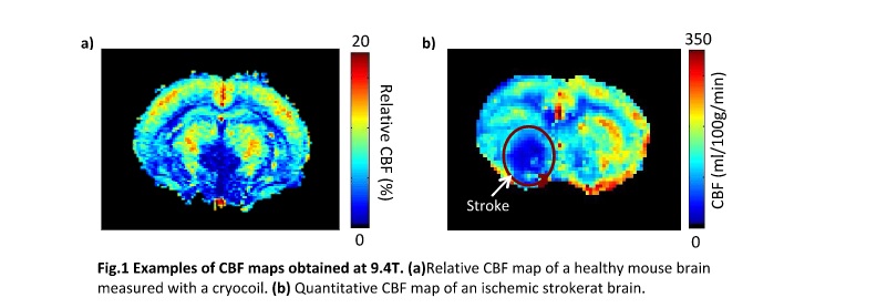
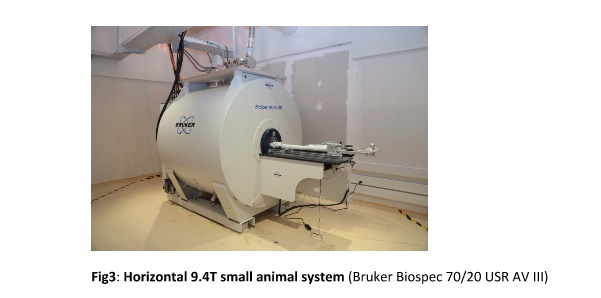
However, quantitative CBF mapping requires several acquisition and post-processing steps that currently limit the use of ASL on preclinical scanners. A user friendly workflow has been developed in collaboration with the scanner constructor Bruker to simplify CBF mapping. CBF maps of rat and mouse brains were acquired at several magnetic fields with different coil setups (eg. at 9.4T: with/without ASL coil, mouse cryocoil) and ASL sequences (CASL, pCASL) on healthy and diseased animals.
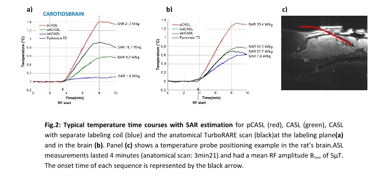
In ASL, the labeling process can lead to heating, especially at higher magnetic fields. This should be taken into account when using ASL for long scanning periods or with diseased animals such as stroke or trauma. To characterize these temperature issues in animals, the specific absorption rate (SAR) delivered to a rat was measured during ASL measurements. Two different labeling sequences (CASL, pCASL) were compared and the benefit of a dedicated labeling coil has been evaluated.
 FLI
FLI


