Coordinator : Monique Bernard, DR CNRS, DU UMR 7339
Project manager : Colette Gomis-Mansaly
Local administrative coordination : University of Aix-Marseille Université
Presentation
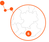
Marseille hub is a multidisciplinary network with high technology imaging equipment and active research and development in methodology and applications in animal models and humans. Pre-clinical and clinical imaging benefit from close connection with research in instrumentation, chemistry and image processing on one side, and research units in biology and clinical departments on the other side. Imaging field has been substantially funded by grants from “Investissements d’avenir” (Infrastructure, Equipex, AMIDEX), ANR, charities (James S McDonnell Foundation, ARSEP, AFM, etc…) and european contracts (M-CUBE FET-Open, IMI pharma-Cog, ERC Gaba Networks, Neuropioneer BrainScale, HBP, Envision, Entervision, Marie Curie, ENDOTOFPET).
All in all, the Marseille hub gathers 200 ETP, including 100 permanent researchers, engineers and clinicians, whose activity on in vivo imaging is partly conducted with prestigious international partners.
The network offers an access to innovative equipment for MRI (at IBISA MR platform of UMR 7339 CRMBM/CEMEREM), nuclear imaging and X ray (at CERIMED, CPPM, APHM, IPC), and optics and biophotonics (at Institut Fresnel, INT, INMED). It includes 3 MRI scanners (4.7T, 7T, 11.75T) for preclinical research, 3 multi-organ MRI systems dedicated to clinical research (1.5T, 3T, 7T), a 3T MRI for preclinical and clinical neuroimaging, 1 clinical PET-CT, 1 clinical SPECT-CT, 1 pre-clinical PET-CT, 1 pre-clinical SPECT-CT, 1 preclinical echograph,1 MEG, 1 bi-photonic spectral imager, one optical non-linear endoscope, one non-linear optical microscope, 1 pre-clinical SPCCT scanner.
The main research topics focus on
-
Innovative advanced optic systems for deep brain imaging in living mouse and the study of myelin sheet organization in mice spinal cord using coherent Raman imaging
-
Ultra-high field MRI in humans (7T) for brain, muscle and osteo-articular imaging, radiofrequency coils with new materials UHF MRI
-
The development of Sodium imaging as an early marker in pathologies (Prof L Schad, Manheim and Dr M Inglese, New-York)
-
Preclinical and clinical neuro–functional MRI
-
Simultaneous EEG and MEG acquisition
-
An X-ray spectral scanner built by Marseille centre of particle physics and based on a new pixel hybrid detector to develop k-edge imaging of Gadolinium and gold nanoparticle. The system will allow dose reduction and molecular imaging thanks to the incorporation of tracer labelled with gadolinium or with gold. This system is installed at CERIMED since the end of 2018.
-
Multimodal brain imaging using simultaneous Magnetoencephalography (MEG), Electroencephalography (EEG) and stereotactic EEG (SEEG) (INS)
Laboratories
CRMBM, CERIMED, Institut Fresnel, INMED, CPPM, INT, LIS
Preclinical and clinical platforms / Managers
Center for Magnetic Resonance in Biology and Medicine/ metabolic exploration by magnetic resonance: CRMBM/CEMEREM UMR7339 – Monique Bernard
European Center for Research in Medical Imaging: Cerimed – Benjamin Guillet
Neurosciences Institute of Timone/fMRI, optics : INT UMR 7289, fMRI manager Jean Luc Anton, photonic imaging manager Ivo Vanzetta
Systems Neurosciences Institute / MEG lab: INS UMR 1106, MEG lab – Jean Michel Badier
Fresnel Institute / Optics, Photonics, Electromagnetism and Signal and Image Processing, UMR 7249: optics and photonics manager, Hervé Rigneault
 FLI
FLI

